Opera Medica et Physiologica

A Cytochemical Study of Iron-Containing Intranuclear Structures of the Human Substantia Nigra Dopaminergic Neurons with Special Emphasis on the Marinesco Bodies

Author Affiliations

Dmitrii E. Korzhevskii *, Elena G. Sukhorukova, Olga V. Kirik, Igor P. Grigorev

Laboratory of Functional Morphology of the Central and Peripheral Nervous System, Department of General and Specific Morphology, Institute of Experimental Medicine, St. Petersburg, 197376 Russia 

Corresponding author: 

Dmitrii E. Korzhevskii (dek2@yandex.ru)

Published ahead of print December 18, 2017; Printed December 25, 2017; OM&P 2017 Volume 3 Issue 3, 4, pages 99-107; doi:10.20388/omp2017.003.0052
Abstract: 

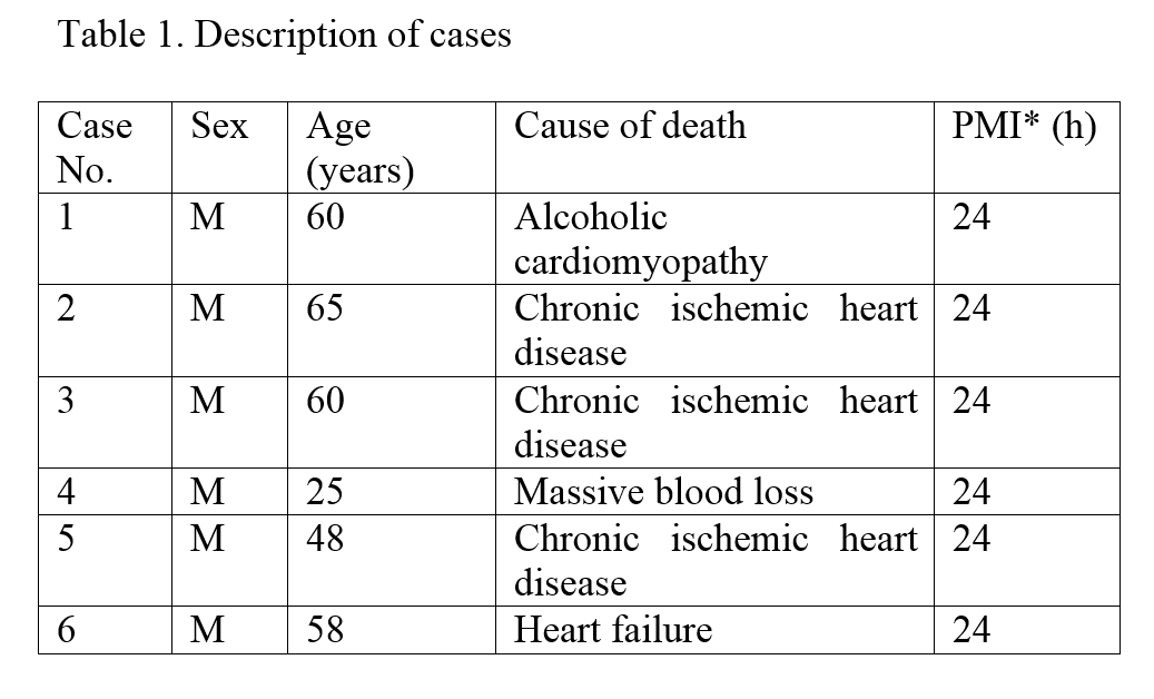
Neurons of the substantia nigra are most prone to degeneration in Parkinson’s disease. The cause of their vulnerability remains unclear and knowledge of the molecular and microstructural features of the substantia nigra pars compacta will help understanding why nigral neurons are vulnerable to damaging factors. 
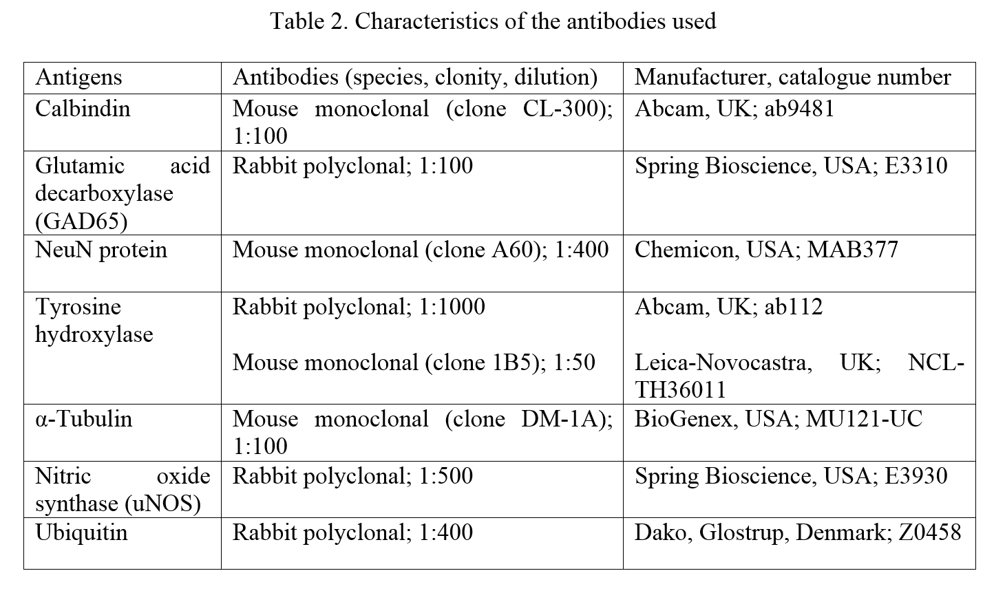
The present study was aimed to investigate the intranuclear inclusions of the nigral neurons, the Marinesco bodies and the Roncoroni rodlets, which origin and function are uncertain, using ubiquitin-, tyrosine hydroxylase-, nitric oxide synthase-, calbindin-, NeuN-, glutamic acid decarboxylase-, and  α-tubulin-immunohistochemistry and iron histochemistry with DAB enhancement.
Of the tested substances, tyrosine hydroxylase and nitric oxide synthase were revealed for the first time in the Marinesco bodies. Non-heme iron was found for the first time in both the Marinesco bodies and the Roncoroni rodlets. In accordance with previous studies, ubiquitin-immunoreactivity was demonstrated in the Marinesco bodies. Moreover, we describe some smaller round and dot-like ubiquitin-immunoreactive structures in the nucleus of melanized neurons. The found small ubiquitin-immunopositive structures within the nucleus are proposed to be the developmental stages of growing Marinesco bodies, whereas Marinesco bodies themselves seem to label the neurons with impaired function of proteasome. 

The substantia nigra is the midbrain area consisting of two parts: the pars compacta and the pars reticulata. The neurons of the pars compacta are mostly dopaminergic with a small portion of glutamatergic cells, whereas those of the pars reticulata are predominantly GABAergic (Nair-Roberts et al., 2008). The dopaminergic nigral neurons are most prone to degeneration in Parkinson’s disease and very vulnerable to various neurotoxins. The cause of their increased vulnerability remains unclear and knowledge of the molecular and microstructural features of the substantia nigra pars compacta will help understanding why these nigral neurons are vulnerable to damaging factors.
Characteristic for the substantia nigra is high level of non-heme iron and iron-containing peptides (Beard et al., 1993; Benkovic & Connor, 1993; Connor et al., 2001; Qian & Wang, 1998; Wang et al., 2007). Iron is an essential trace element required for synthesis and metabolism of some neurotransmitters (including dopamine), formation of myelin, transport of oxygen, and oxidative metabolism in mitochondria (Glinka et al., 1997; Youdim et al., 1991). A number of studies demonstrated that Parkinson’s disease is associated with elevated iron levels in the substantia nigra pars compacta (Andersen, 2004; Jin et al., 2012; Lan & Jiang, 1997; Lv et al., 2011; Sofic et al., 1991) and, moreover, in individual nigral dopaminergic neurons (Oakley et al., 2007). These observations gave rise to the proposal that iron and iron-induced oxidative stress have a causative role in progressive neurodegeneration of the substantia nigra neurons in Parkinson’s disease (Andersen, 2004; Benarroch, 2009; Berg et al., 2008; Götz et al., 2004; Kaur & Andersen, 2004; Ke & Ming Qian, 2003; Medeiros et al., 2016; Sian-Hülsmann et al., 2011).
Another known distinctive feature of the human substantia nigra is a pigment neuromelanin in the cytoplasm of the dopaminergic nerve cells (In fact, not all dopaminergic neurons have melanin and not all melanin-containing neurons are dopaminergic (Grigoriev et al., 2013; Sukhorukova et al., 2014)). Origin, composition and function(s) of neuromelanin are not fully understood despite intensive investigations for decades (D’Ischia & Prota, 1997; Engelen et al., 2012; Zecca et al., 1996; Zucca et al., 2015). Of interest is that neuromelanin in nigral neurons was shown to contain iron (Double et al., 2003; Jellinger et al., 1992) and it seems just this iron is responsible for induction of the oxidative stress resulting in neurodegeneration of nigral neurons in Parkinson’s disease (Ben-Shachar et al., 1991; Faucheux et al., 2003; Shamoto-Nagai et al., 2006).
A unique property of the melanin-containing neurons of the human substantia nigra is occurrence of the so-called Marinesco bodies in their nucleus. These eosinophilic spherical intranuclear inclusions have been described for the first time in 1902 by G. Marinesco. More than a hundred years later, the origin and molecular composition of this intranuclear structure are not clearly understood, while its function(s) is enigmatic. Structure of the Marinesco bodies has been studied using light microscopy (Anraku et al., 1970; Yuen & Baxter, 1963), histochemistry (Yuen & Baxter, 1963), electron microscopy (Leestma & Andrews, 1969; Okamoto & Hirai, 1981), and, more recently, with immunohistochemistry. Immunohistochemical investigations of the Marinesco bodies revealed ubiquitin and some other proteins implicated in the ubiquitin–proteasome system and autophagy (Dickson et al., 1990; Woulfe et al., 2004; Odagiri et al., 2012; Mori et al., 2012) as well as some other polypeptides (Mori et al., 2012b; Mizuno et al., 2003). Although Marinesco bodies were found in the substantia nigra in some neurodegenerative diseases (Anraku et al., 1970; Mizuno et al., 2003; Mori et al., 2012a,b; Takahashi-Fujigasaki & Fujigasaki, 2006) no interdependence has been established between the presence of the Marinesco bodies in nucleus of the nigral neurons and any neuropathological state. The only correlation was found between the frequency of the Marinesco bodies and the age of the human: the older the human, the more Marinesco bodies can be found in the nucleus of the nigral neurons (Dickson et al., 1990; Alladi et al., 2010). 
Another intranuclear inclusion, the Roncoroni rodlet can also be observed in the nigral neurons. The intranuclear rod-like inclusions have been discovered in nerve cells in 1890s (Mann, 1894; Roncoroni, 1895) and Ramon y Cajal referred to it as the rodlet of Roncoroni (Ramon y Cajal, 1909, 1911). Recently, Roncoroni rodlets were shown to express tubulin, glucocorticoid receptor-like, and promyelocytic leukaemia immunoreactivity (Woulfe & Munoz, 2000; Woulfe et al., 2002, 2007). A marked reduction of Roncoroni rodlets in Alzheimer's disease brain was the only correlation reported between these intranuclear inclusions and the brain pathology (Woulfe et al., 2002). However, composition of these microstructures has not yet been completely clarified. Similarly to the Marinesco bodies, exact function(s) of Roncoroni rodlets remains unknown. 
The above mentioned facts show some unique features of the substantia nigra each of which can contribute to high vulnerability of the nigral dopaminergic melanin-containing neurons. The present study was aimed to investigate the subcellular distribution of non-heme iron and some proteins in the neurons of the human substantia nigra pars compacta using immunohistochemistry and iron histochemistry with emphasis on the intranuclear inclusions. Immunohistochemical visualization of tyrosine hydroxylase, the key enzyme of dopamine synthesis, was used to check the preservation of the nigral tissue as a whole and, particularly, nigral dopaminergic neurons, and to define the boundaries of the substantia nigra pars compacta.
 
Materials and Methods
 
Tissue preparation
 
Adult human brain samples were collected from neurologically normal individuals submitted to autopsy (see Table 1 for description of cases). Brain tissue blocks (approx. 10x10x10-mm) containing the substantia nigra were fixed in the zinc-ethanol-formalin fixative (ZEF) prepared according to previously published protocol (Korzhevskii et al., 2014; Korzhevskii et al., 2015): 1 g ZnCl2 was dissolved in mixture of 90 ml 96 % ethanol and 10 ml concentrated (35-39 %) formaldehyde (all purchased from Vekton, St. Petersburg, Russia). The tissue blocks were allowed to stay in the fixative for 24 hours at room temperature, dehydrated, and embedded in paraffin routinely. Sections 7 µm thick were cut using sliding microtome (Leica SM 2000R, Leica, Germany) and mounted on silane coated glass slides (HistoBondR, Marienfeld, Germany). 
The study project was positively approved by the local Ethics Committee of the Institute of Experimental Medicine, (St. Petersburg, Russia).
A part of the sections were Nissl stained with either cresyl violet (cresyl violet for microscopy, Merck, Darmstadt, Germany), or toluidine blue O (Acros Organics, Geel, Belgium).
 
Immunohistochemistry
 
Ubiquitin, tyrosine hydroxylase, calbindin, glutamic acid decarboxylase, NeuN, α-tubulin, and nitric oxide synthase immunohistochemistry was performed according to previously used protocols (Korzhevskii et al., 2015, 2017; Grigoriev et al., 2012).
For immunohistochemistry the sections were deparaffinized and rehydrated routinely. The heat-induced antigen retrieval was performed immunohistochemistry as follows: the sections were incubated in modified citrate buffer, pH 6.1 (S1700, Dako, Glostrup, Denmark) in a conventional steamer for 25 min. The preparations were pretreated with the blocking solution (Protein Block, Spring Bioscience, Pleasanton, CA, USA) and then, a primary antibody was applied (for details see Table 2). 
For visualization of the primary antibodies, the HRP-conjugate from the Reveal Polyvalent HRP DAB Detection System (Spring Bioscience, Pleasanton, CA, USA) was applied for rabbit polyclonal antibodies and EnVision+ System-HRP Labelled Polymer Anti-Mouse (Dako, Denmark) or MACH2 Mouse HRP-Polymer (Biocare Medical, USA) – for mouse monoclonal antibodies. Immunostained sections were counterstained with either cresyl violet (cresyl violet for microscopy, Merck, Darmstadt, Germany) or alum hematoxylin.
Control of the immunohistochemical reaction was performed according to the recommendation of the reagent manufacturers. 
 
Iron histochemistry (Perls’ reaction). 
 
After deparaffinization and rehydration, the sections were treated with 3 % hydrogen peroxide for 10 min, then, washed in distilled water for 3-5 min, and incubated in freshly prepared mixture of 2 % aqueous  solution of potassium ferrocyanide  (K4[Fe(CN)6] · 3H2O) and 0.2 N hydrochloric acid (1 : 1) for 30 min at room temperature. This protocol is consistent with the standard Perls’ reaction and gives blue-coloration of the non-heme iron accumulations. 
Some of the preparations were examined after the standard Perls’ reaction, while others were subjected to intensification of the Perls’ reaction according to Nguyen-Legros et al. (Nguyen-Legros et al., 1980) in Meguro modification [Meguro et al., 2007) as described in details elsewhere (Sukhorukova et al., 2013). Briefly, after the standard Perls’ reaction, the sections were thoroughly washed in distilled water (3 portions for 3-5 min) and, then, treated with the chromogen 3,3'-diaminobenzidine (DAB) (Dako, Denmark) for 10-15 min. The exact time of treatment was adjusted according to development of optimal staining with DAB in the control preparations knowingly containing Fe3+. Finally, the preparations were counterstained with 0.5% nuclear fast red (Sigma-Aldrich, USA) prepared as follows: 1 g nuclear fast red and 50 g potassium aluminum sulfate was dissolved in 500 ml distilled water.
 
Light microscopy
 
The preparations were examined with a Leica DM 750 microscope and photographed using a Leica ICC 50 digital camera (Leica Microsystems, Wetzlar, Germany) operated by LAS EZ software (ver. 1.8.0, Leica Microsystems, Heerbrugg, Switzerland).
 
Figure 1. Marinesco body-like round structure in the nucleus of the melanized neuron in the human substantia nigra pars compacta. Nissl-staining with toluidine blue. Single arrow – weakly stained Marinesco body-like structure with a pale halo in the neuronal nucleus; double arrow – nucleolus, asterisk (*) – neuromelanin. Bar scale – 20 µm.
Figure 1. Marinesco body-like round structure in the nucleus of the melanized neuron in the human substantia nigra pars compacta. Nissl-staining with toluidine blue. Single arrow – weakly stained Marinesco body-like structure with a pale halo in the neuronal nucleus; double arrow – nucleolus, asterisk (*) – neuromelanin. Bar scale – 20 µm.
 
Results
 
Nissl stained sections demonstrated good preservation of all studied specimens of the human substantia nigra (Fig. 1) and the tyrosine hydroxylase-immunohistochemistry showed normal appearance of the dopaminergic neurons in the substantia nigra pars compacta (data not shown), most of which demonstrated presence of high amounts of neuromelanin granules (Fig. 1, 2, 3). Many neurons has round intranuclear inclusions (more often 2-4 in number) of different sizes varying from small (much smaller than the nucleolus) to nucleolus-large spheres, resembling the Marinesco bodies. Frequency of their appearance in the neuronal nucleus varied significantly among the substantia nigra specimens from different brains. Staining with the toluidine blue clearly distinguishes the nucleolus from these intranuclear inclusions: the nucleolus is stained bright blue in contrast to weakly stainable round intranuclear inclusions (Fig. 1). Generally, the weakly stained round intranuclear inclusion has a halo of almost unstained material (Fig. 1).
Figure 2. Ubiquitin immunohistochemistry of the human substantia nigra pars compacta. Asterisk (*) indicates neuromelanin accumulation in the neuronal cytoplasm; single arrow – ubiquitin-immunopositive (brown) structures in the neuronal nucleus; double arrow – nucleolus. Note the weak ubiquitin immunoreactivity in the nucleoplasm (B). Counterstained with cresyl violet. Bar scale – 10 µm.
Figure 2. Ubiquitin immunohistochemistry of the human substantia nigra pars compacta. Asterisk (*) indicates neuromelanin accumulation in the neuronal cytoplasm; single arrow – ubiquitin-immunopositive (brown) structures in the neuronal nucleus; double arrow – nucleolus. Note the weak ubiquitin immunoreactivity in the nucleoplasm (B). Counterstained with cresyl violet. Bar scale – 10 µm. 
 
 
In some neuronal nuclei, an elongated rod-shaped inclusion was seen. As a rule, these rodlets were laying in close proximity to the nucleolus and they have nearly the same length as the diameter of the nucleus. None of neuronal nucleus contained the round and the rod-like intranuclear inclusions together.
Ubiquitin immunohistochemistry revealed the ubiquitin in the round intranuclear inclusions of nigral neurons (Fig. 2A,B). All the found round intranuclear inclusions irrespectively their sizes were ubiquitin-immunoreactive. Moreover, the ubiquitin-immunoreactivity was found in some small roundish and dot-like structures that were much smaller than the common round intranuclear inclusions (Fig. 2B). It should be noted that not all nuclei contain the round intranuclear inclusions, but all (or at least most of) the round intranuclear inclusion-containing nuclei exhibit weak ubiquitin immunoreactivity in the nucleoplasm. 
Iron histochemistry (standard Perls’ reaction) demonstrated a prussian blue deposit in many nigral cells of various origin: microglia- and oligodendroglia-like cells, in the perivascular cells, and in the substantia nigra parenchima (Fig. 3A). However, standard iron histochemistry failed to stain nigral neurons. DAB enhancing technique gives more intensive homogeneous staining of the pars compacta of the substantia nigra. In this case, the product of the histochemical reaction was revealed in neurons, glia, and some non-identifiable structures of the substantia nigra. In the neurons, the iron deposits were observed in clumps of neuromelanin and the intranuclear round and rod-like inclusions which morphological appearance corresponds to Marinesco bodies and Roncoroni rodlets, respectively (Fig. 3B,C). Not all inclusions were histochemically stained. The intranuclear rodlets have normally a deposit of the histochemical reaction along the entire length of the rods.
Figure 3. Perls’ histochemical reaction for iron in the human substantia nigra pars compacta.A) standard Perls’ reaction without DAB enhancement. Single arrow – granular cytoplasmic inclusions with iron-positive reaction (blue) in a perivascular cell; B) and C)  Perls’ reaction with DAB enhancement. Single arrow – iron-positive (brown) round intranuclear inclusion (B), iron-positive (brown) rod-like intranuclear inclusion (C). Note the iron-positive reaction of neuromelanin after usage of DAB enhancement technique (B and C). Asterisk (*) indicates neuromelanin accumulation in the neuronal cytoplasm (A-C); double arrow – nucleolus. Counterstaining with the nuclear fast red (A-C). Bar scale – 20 µm.
Figure 3. Perls’ histochemical reaction for iron in the human substantia nigra pars compacta.A) standard Perls’ reaction without DAB enhancement. Single arrow – granular cytoplasmic inclusions with iron-positive reaction (blue) in a perivascular cell; B) and C)  Perls’ reaction with DAB enhancement. Single arrow – iron-positive (brown) round intranuclear inclusion (B), iron-positive (brown) rod-like intranuclear inclusion (C). Note the iron-positive reaction of neuromelanin after usage of DAB enhancement technique (B and C). Asterisk (*) indicates neuromelanin accumulation in the neuronal cytoplasm (A-C); double arrow – nucleolus. Counterstaining with the nuclear fast red (A-C). Bar scale – 20 µm.


Special immunohistochemical investigation of intranuclear inclusions with a number of antigens (Table 2) revealed tyrosine hydroxylase and nitric oxide synthase staining in the Marinesco bodies (Fig. 4A,B). Tyrosine hydroxylase immunoreactivity was homogeneous and distinctly visible in some round intranuclear inclusions, that were less in diameter than the nucleolus and non-stained with hematoxyline. In some melanin-containing neurons, the nitric oxide synthase immunoreactivity was evident in small Marinesco body-like round intranuclear inclusions with dense staining of the core and pale thin perimembranous halo. Noteworthy, the round intranuclear inclusions stained for both tyrosine hydroxylase and nitric oxide synthase were intensively immunoreactive in contrast to other compartments of the nucleus with weak (if any) immunostaining.  
Figure 4. Marinesco bodies in melanized neurons of the human substantia nigra pars compacta. A) Intensive tyrosine hydroxylase-immunoreactivity (brown) is distributed mostly in the cytoplasm of a neuron and many processes. The neuronal nucleus exhibits weak tyrosine hydroxylase-immunoreactivity with exception of a pronounced immunohistochemical reaction in the round Marinesco body-like structure (single arrow). Nucleolus (double arrow) is devoid of tyrosine hydroxylase-immunoreactivity. Asterisk (*) indicates neuromelanin accumulation in the neuronal cytoplasm. B) Intensive nitric oxide synthase-immunoreactivity (brown) is seen predominantly in the cytoplasm of a neuron with many neuromelanin granules (asterisk). The neuronal nucleus is mostly immunonegative with only one nitric oxide synthase-immunoreactive round Marinesco body-like structure (single arrow). Note a homogeneously stained core and a pale perimembranous halo. Double arrow indicates nucleolus. Counterstaining with hematoxylin. Bar scale – 10 µm.
Figure 4. Marinesco bodies in melanized neurons of the human substantia nigra pars compacta. A) Intensive tyrosine hydroxylase-immunoreactivity (brown) is distributed mostly in the cytoplasm of a neuron and many processes. The neuronal nucleus exhibits weak tyrosine hydroxylase-immunoreactivity with exception of a pronounced immunohistochemical reaction in the round Marinesco body-like structure (single arrow). Nucleolus (double arrow) is devoid of tyrosine hydroxylase-immunoreactivity. Asterisk (*) indicates neuromelanin accumulation in the neuronal cytoplasm. B) Intensive nitric oxide synthase-immunoreactivity (brown) is seen predominantly in the cytoplasm of a neuron with many neuromelanin granules (asterisk). The neuronal nucleus is mostly immunonegative with only one nitric oxide synthase-immunoreactive round Marinesco body-like structure (single arrow). Note a homogeneously stained core and a pale perimembranous halo. Double arrow indicates nucleolus. Counterstaining with hematoxylin. Bar scale – 10 µm. 
 
 
Discussion
 
Previous investigation of the human substantia nigra specimens fixed in the zinc-ethanol-formalin fixative demonstrated good preservation of the tissue, which is a prerequisite to adequate results in further immunohistochemical and histochemical studies (Korzhevskii et al., 2015). Good preservation of the human substantia nigra tissue after fixation in zinc-ethanol-formaldehyde was confirmed in the present study using both Nissl staining and tyrosine hydroxylase-immunohistochemistry. The latter was used as a standard immunohistochemical staining for the substantia nigra cells and demonstrated normal appearance of the nigral dopaminergic neurons. 
The round intranuclear inclusions revealed in the melanized nigral neurons are consistent with the Marinesco bodies described as early as 1902 (Marinesco, 1902). However, after a century of studying, the origin, molecular composition, and the function(s) of these intranuclear structures remain to be elucidated. Therefore, studies of the chemical composition of this intranuclear inclusion brings us closer to understanding its significance. Previous immunohistochemical investigations has demonstrated that ubiquitin is an obligatory component of the Marinesco bodies (Dickson et al., 1990; Woulfe et al., 2004; Odagiri et al., 2012), whereas a number of some other proteins may be contained in these structures as well (Odagiri et al., 2012; Mori et al., 2012a, b; Mizuno et al., 2003; Takahashi-Fujigasaki & Fujigasaki, 2006; Alladi et al., 2010). In the present study, we also observed ubiquitin in the Marinesco bodies. Furthermore, ubiquitin was found not in large (“mature”) Marinesco bodies only, but also in some smaller intranuclear roundish and dot-like structures suggesting that these ubiquitin-immunoreactive structures are putative predecessors of the Marinesco bodies and the latter are formed from an ubiquitin-containing matter from the very beginning. Ubiquitin as a component of ubiquitin-proteasome system marks the aberrant proteins for degradation (Hershko & Ciechanover, 2005). Therefore, similar to cytoplasm, the ubiquitin in the nucleus is most likely present in the form of conjugate with the damaged proteins that were not destructed via the proteasome for some reason. If so, the Marinesco bodies are largely composed of damaged proteins not subjected to destruction by proteasome and are seen in and mark the nucleus of neurons with impaired proteasome function. The aging was shown to be associated with decreased proteasome activity and accumulation of ubiquitinated proteins (Tsakiri & Trougakos, 2015). This is in parallel with an increase in number of the Marinesco bodies (consisting of ubiquitinated proteins) in nucleus of nigral neurons with aging and can be regarded as a reflection of decreased proteasome activity in these cells. In this context, the found weak ubiquitin immunoreactivity over the nucleoplasm of the Marinesco body-containing nuclei seems to be a result of decreased function of nuclear proteasome and, accordingly, accumulation of dispersed ubiquitin-tagged aberrant proteins in the nucleus as an initial step toward formation of the Marinesco bodies.
Present study is first to demonstrate tyrosine hydroxylase and nitric oxide synthase in the Marinesco body. Tyrosine hydroxylase is a well-known and widely used marker of the catecholaminergic neurons, including dopaminergic neurons of the substantia nigra and nitric oxide synthase has also been shown in the nigral neurons (Korzhevskii et al., 2017; Cavalcanti-Kwiatkoski et al., 2010; Czarnecka et al., 2013; González-Hernández et al., 1997; Tan et al., 2016). Although both tyrosine hydroxylase and nitric oxide synthase are predominantly cytoplasmic enzymes, they are expressed to a lesser extent in nucleus as well, but has heretofore not been shown in the Marinesco bodies. As the Marinesco body, according to the above mentioned proposal, consists of ubiquitin-tagged aberrant proteins, appearance of common nigral proteins the tyrosine hydroxylase and the nitric oxide synthase in the Marinesco bodies is quite probable. However, why these cytoplasmic enzymes, irrespectively unbound (normal or defective), or ubiquitin-tagged (defective) enter the nucleus and accumulate there in the form of Marinesco body remains elusive.
Iron is essential trace element involved in numerous redox reactions in the nervous system (Shinobu & Beal, 1997). Moreover, iron-containing proteins has been shown in the nucleus (Cheepsunthorn et al., 1998; Geuens et al., 2003; Korzhevskii et al., 2015), where they contribute to DNA replication and repair (Wu & Brosh, 2012). However, distribution of the iron inside the nucleus has not yet been studied. 
In the present study, we used the Perls’ method for ferric iron to reveal localization of non-heme iron in the nigral cells. Although Perls’ method is one of the oldest histochemical technique, it is successfully used for nervous and non-nervous tissues in animal and human studies (Benkovic & Connor, 1993; Dwork et al., 1988; Grizzi et al., 2002; Kondo et al., 1995; Li et al., 2015; Vidal et al., 2008). Standard Perl’s staining visualized ferrous iron in the substantia nigra, but failed to show it in neurons of the substantia nigra, which is in agreement with previous data (Yuen & Baxter,  1963). However, use of DAB-enhanced iron histochemistry allowed demonstration of non-heme iron in the nigral neurons. In the cytoplasm, the iron was observed in the neuromelanin aggregates, which is consistent with the studies that have showed iron-neuromelanin interaction in the substantia nigra (Faucheux et al., 2003; Zecca et al., 2004; Dwork et al., 1988; Double et al., 2003; Jellinger et al., 1992; Zucca et al., 2015). The non-heme iron in nucleus of neurons was found recently (Sukhorukova et al., 2013), and the present study demonstrates for the first time the non-heme iron in the Marinesco bodies and the Roncoroni rodlets. Finding of the non-heme iron in these intranuclear inclusions apparently indicates the presence of iron-containing proteins in these intranuclear inclusions. The origin and function of both intranuclear inclusions is yet uncertain and finding of iron in this structure is a step toward a better understanding of the chemical composition and physiological role of these intranuclear structures.
In conclusion, the present study demonstrates occurrence of tyrosine hydroxylase and nitric oxide synthase in the Marinesco bodies of the melanin-containing nigral neurons. Nucleus of many nigral dopaminergic neurons has one or several typical ubiquitin-immunoreactive Marinesco bodies and a lot of smaller rounded and dot-like ubiquitin-immunoreactive microstructures, that are proposed to be the predecessors of the “mature” Marinesco bodies. Moreover, the formation of the Marinesco bodies seems to be associated with accumulation of ubiquitin-tagged aberrant proteins in the nucleus because of aging-related proteasome functional insufficiency in nigral neurons. Consequently, the Marinesco bodies label the nigral neurons with aging-related impairment of proteasome function. 
Both the Marinesco bodies and the Roncoroni rodlets were shown to contain non-heme iron and, thus, can be involved in normal or aberrant metabolism of the iron-containing proteins in the nucleus of nigral neurons.  
 
Conflict of interests: the authors declare that there is no conflict of interest that could be perceived as prejudicing the impartiality of the research reported.
 
 
AttachmentSize
omp2017.003.0052.pdf6.15 MB
References: 

ALLADI P.A., MAHADEVAN A., VIJAYALAKSHMI K., MUTHANE U., SHANKAR S.K. & RAJU T.R. (2010): Ageing enhances alpha-synuclein, ubiquitin and endoplasmic reticular stress protein expression in the nigral neurons of Asian Indians. Neurochemistry International 57, 530-539.
ANDERSEN J.K. (2004): Iron dysregulation and Parkinson's disease. Journal of Alzheimer's Disease 6, S47-52. 
ANRAKU S., MIURA C., MORI H. & FUKUTOMI T. (1970): Histological studies on the intranuclear inclusions of the melanin-pigmented nerve cells in the substantia nigra and the nucleus loci caerulei. Kurume Medical Journal 17, 211-224.
BEARD J. L., CONNOR J. R. & JONES B. C. (1993): Brain iron: location and function. Progress in Food & Nutrition Science 17, 183–221.
BENKOVIC S.A. & CONNOR J.R. (1993): Ferritin, transferrin, and iron in selected regions of the adult and aged rat brain. Journal of Comparative Neurology 338, 97-113.
BENARROCH E.E. (2009): Brain iron homeostasis and neurodegenerative disease. Neurology 72, 1436-1440.
BEN-SHACHAR D., RIEDERER P. & YOUDIM M.B. (1991): Iron-melanin interaction and lipid peroxidation: implications for Parkinson's disease. Journal of Neurochemistry 57, 1609-1614.
BERG D., RIEDERER P. & GERLACH M. (2008): Contribution of disturbed iron metabolism to the pathogenesis of Parkinson’s disease. Future Neurology 3, 447-461.
CAVALCANTI-KWIATKOSKI R., RAISMAN-VOZARI R., GINESTET L. & DEL BEL E. (2010): Altered expression of neuronal nitric oxide synthase in weaver mutant mice. Brain Research 1326, 40-50.
CHEEPSUNTHORN P., PALMER C. & CONNOR J.R. (1998): Cellular distribution of ferritin subunits in postnatal rat brain. Journal of Comparative Neurology 400, 73–86. 
CONNOR J.R., MENZIES S.L., BURDO J.R. & BOYER P.J. (2001): Iron and iron management proteins in neurobiology. Pediatric Neurology 2001; 25, 118-129.
CZARNECKA A., LENDA T., DOMIN H., KONIECZNY J., SMIALOWSKA M. & LORENC-KOCI E. (2013): Alterations in the expression of nNOS in the substantia nigra and subthalamic nucleus of 6-OHDA-lesioned rats: the effects of chronic treatment with l-DOPA and the nitric oxide donor, molsidomine. Brain Research 1541, 92-105.
DICKSON D.W., WERTKIN A., KRESS Y., KSIEZAK-REDING H. & YEN S.H. (1990): Ubiquitin immunoreactive structures in normal human brains. Distribution and developmental aspects. Laboratory Investigation 63, 87-99. 
D’ISCHIA M. & PROTA G. (1997): Biosynthesis, structure, and function of neuromelanin and its relation to Parkinson’s disease: A Critical Update. Pigment Cell Research 10, 470-476.
DOUBLE K.L., GERLACH M., SCHÜNEMANN V., TRAUTWEIN A.X., ZECCA L., GALLORINI M., YOUDIM M.B., RIEDERER P. & BEN-SHACHAR D. (2003): Iron-binding characteristics of neuromelanin of the human substantia nigra. Biochemical Pharmacology 66, 489-494.
DWORK A.J., SCHON E.A. & HERBERT.J. (1988): Nonidentical distribution of transferrin and ferric iron in human brain. Neuroscience 27, 333-345.
ENGELEN M., VANNA R., BELLEI C., ZUCCA F.A., WAKAMATSU K., MONZANI E., ITO S., CASELLA L. & ZECCA L. (2012): Neuromelanins of human brain have soluble and insoluble components with dolichols attached to the melanic structure. Public Library of Science One 7, e48490.
FAUCHEUX B.A., MARTIN M.E., BEAUMONT C., HAUW J.J., AGID Y. & HIRSCH E.C. (2003): Neuromelanin associated redox-active iron is increased in the substantia nigra of patients with Parkinson's disease. Journal of Neurochemistry 86, 1142-1148.
GEUENS E., BROUNS I., FLAMEZ D., DEWILDE S., TIMMERMANS J.P. & MOENS L. (2003): A globin in the nucleus! Journal of Biological Chemistry 278, 30417-30420. 
GLINKA Y., GASSEN M. & YOUDIM M.B.H. (1997): Iron and neurotransmitter function in the brain. In: Metals and Oxidative Damage in Neurological Disorders (Ed Connor J.R.), pp. 1-22, New York: Springer Science Media.
GONZÁLEZ-HERNÁNDEZ T., ABDALA P.  & RODRÍGUEZ M. (1997): NOS expression in nigral cells after excitotoxic and non-excitotoxic lesion of the pedunculopontine tegmental nucleus. European Journal of Neuroscience 9, 2658-2667.
GÖTZ M.E., DOUBLE K., GERLACH M., YOUDIM M.B. & RIEDERER P. (2004): The relevance of iron in the pathogenesis of Parkinson's disease. Annals of the New York Academy of Sciences 1012, 193-208.
GRIGORIEV I.P., SUKHORUKOVA E.G., KOLOS E.A. & KORZHEVSKII D.E. (2013): Neuromelanin in substantia nigra neurons lacking tyrosine hydroxylase. Neuroscience and Behavioral Physiology 43, 461-463.
GRIGORIEV I.P., VASILENKO M.S., SUKHORUKOVA E.G. & KORZHEVSKII D.E. (2012): Use of different antibodies to tyrosine hydroxylase to study catecholaminergic systems in the mammalian brain. Neuroscience and Behavioral Physiology 42, 210-213.
GRIZZI F., CEVA-GRIMALDI G., FRANCESCHINI B., RONCALLI M., CHIRIVA-INTERNATI M. & DIOGUARDI N. (2002): Simultaneous staining of cytoplasmic iron and collagen matrix in human liver biopsy specimens. European Journal of Histochemistry 46, 101-104.
HERSHKO A. & CIECHANOVER A. (1998): The ubiquitin system. Annual Review of Biochemistry 67, 425–79. 
JELLINGER K., KIENZL E., RUMPELMAIR G., RIEDERER P., STACHELBERGER H., BEN-SHACHAR D. & YOUDIM M.B. (1992): Iron-melanin complex in substantia nigra of parkinsonian brains: an x-ray microanalysis. Journal of Neurochemistry; 59, 1168-1171.
JIN L., WANG J., JIN H., FEI G., ZHANG Y., CHEN W., ZHAO L., ZHAO N., SUN X., ZENG M. & ZHONG C. (2012): Nigral iron deposition occurs across motor phenotypes of Parkinson's disease. European Journal of Neurology 19, 969-976.
KAUR D. & ANDERSEN J. (2004): Does cellular iron dysregulation play a causative role in Parkinson's disease? Ageing Research Reviews 3, 327-343.
KE Y. & MING QIAN Z. (2003): Iron misregulation in the brain: a primary cause of neurodegenerative disorders. The Lancet Neurology 2, 246-253.
KONDO Y., OGAWA N., ASANUMA M., OTA Z. & MORI A. (1995): Regional differences in late-onset iron deposition, ferritin, transferrin, astrocyte proliferation, and microglial activation after transient forebrain ischemia in rat brain. Journal of Cerebral Blood Flow & Metabolism 15, 216-226.
KORZHEVSKII D.E., GRIGOR’EV I.P., KIRIK O.V. & ALEKSEEVA O.S. (2015): Neuroglobin distribution in the rat cerebellar Purkinje cells. Journal of Evolutionary Biochemistry and Physiology 51, 517-519.
KORZHEVSKII D.E., GRIGOR'EV I.P., SUKHORUKOVA E.G. & GUSEL'NIKOVA V.V. (2017): [Immunohistochemical characteristics of the substantia nigra neurons of the human]. Russian. Zhurnal Nevrologii i Psikhiatrii imeni S.S. Korsakova 117, 50-55.
KORZHEVSKII  D.E., SUKHORUKOVA  E.G., GILEROVICH  E.G., PETROVA  E.S., KIRIK O.V. & GRIGOREV  I.P. (2014): Advantages and disadvantages of zinc-ethanol-formaldehyde as a fixative for immunocytochemical studies and confocal laser microscopy. Neuroscience Behavioral Physiology 44, 542-545.
KORZHEVSKII D.E., SUKHORUKOVA E.G., KIRIK O.V. & GRIGOREV I.P. (2015): Immunohistochemical demonstration of specific antigens in the human brain fixed in zinc-ethanol-formaldehyde. European Journal of Histochemistry 59, 2530.
LAN J. & JIANG D.H. (1997): Excessive iron accumulation in the brain: a possible potential risk of neurodegeneration in Parkinson’s disease. Journal of Neural Transmission 104, 649–660. 
LEESTMA J.E. & ANDREWS J.M. (1969): The fine structure of the Marinesco body. Archives of Pathology 88, 431-436. 
LI W., GARRINGER H.J., GOODWIN C.B., RICHINE B., ACTON A., VANDUYN N., MUHOBERAC B.B., IRIMIA-DOMINGUEZ J., CHAN R.J., PEACOCK M., NASS R., GHETTI B. & VIDAL R. (2015): Systemic and cerebral iron homeostasis in ferritin knock-out mice. Public Library of Science One 10, e0117435.
LV Z., JIANG H., XU H., SONG N. & XIE J. (2011): Increased iron levels correlate with the selective nigral dopaminergic neuron degeneration in Parkinson's disease. Journal of  Neural Transmission (Vienna)  118, 361-369.
MANN G. (1894): Histological changes induced in sympathetic, motor, and sensory nerve cells by functional activity (preliminary note). Journal of Anatomy and Physiology 29, 100-108. 
MARINESCO G. (1902): [On the presence of paranucleolar acidophilic corpuscles in the cells of the locus niger and the locus coeruleus. French.] Comptes Rendus de l'Académie des Sciences 135, 1000-1002. 
MEDEIROS M.S., SCHUMACHER-SCHUH A., CARDOSO A.M., BOCHI G.V., BALDISSARELLI J., KEGLER A., SANTANA D., CHAVES C.M., SCHETINGER M.R., MORESCO R.N., RIEDER C.R. & FIGHERA M.R. (2016): Iron and oxidative stress in parkinson’s disease: an observational study of injury biomarkers. Public Library of Science one 11, e0146129.
MEGURO R., ASANO Y., ODAGIRI S., LI C., IWATSUKI H. & SHOUMURA K. (2007): Nonheme-iron histochemistry for light and electron microscopy: a historical, theoretical and technical review. Archives of Histology and Cytology 70, 1-19.
MIZUNO Y., HORI S., KAKIZUKA A. & OKAMOTO K. (2003): Vacuole-creating protein in neurodegenerative diseases in humans. Neuroscience Letters 343, 77-80.
MORI F., TANJI K., ODAGIRI S., TOYOSHIMA Y., YOSHIDA M., KAKITA A., TAKAHASHI H. & WAKABAYASHI K. (2012a): Autophagy-related proteins (p62, NBR1 and LC3) in intranuclear inclusions in neurodegenerative diseases. Neuroscience Letters 522, 134-138.
MORI F., TANJI K., TOYOSHIMA Y., YOSHIDA M., KAKITA A., TAKAHASHI H. & WAKABAYASHI K. (2012b): Optineurin immunoreactivity in neuronal nuclear inclusions of polyglutamine diseases (Huntington's, DRPLA, SCA2, SCA3) and intranuclear inclusion body disease. Acta Neuropathologica 123, 747-749. 
NAIR-ROBERTS R.G., CHATELAIN-BADIE S.D., BENSON E., WHITE-COOPER H., BOLAM J.P. & UNGLESS M.A. (2008): Stereological estimates of dopaminergic, GABAergic and glutamatergic neurons in the ventral tegmental area, substantia nigra and retrorubral field in the rat. Neuroscience 152, 1024-1031.
NGUYEN-LEGROS J., BIZOT J., BOLESSE M. & PULICANI J.P. 1980: ["Diaminobenzidine black" as a new histochemical demonstration of exogenous iron (author's transl). French.]. Histochemistry 66, 239-244. 
OAKLEY A.E., COLLINGWOOD J.F., DOBSON J., LOVE G., PERROTT H.R., EDWARDSON J.A., ELSTNER M. & MORRIS C.M. (2007): Individual dopaminergic neurons show raised iron levels in Parkinson disease. Neurology 68, 1820-1825.
ODAGIRI S., TANJI K., MORI F., KAKITA A., TAKAHASHI H., KAMITANI T. & WAKABAYASHI K. (2012): Immunohistochemical analysis of Marinesco bodies, using antibodies against proteins implicated in the ubiquitin–proteasome system, autophagy and aggresome formation. Neuropathology 32, 261–266. 
OKAMOTO K. & HIRAI S. (1981): [Fine structures of Marinesco body and nuclear body in the substantia nigra. Japanese.] Rinsho Shinkeigaku [Clinical Neurology] 21, 781-789. 
QIAN Z.M. & WANG Q. (1998): Expression of iron transport proteins and excessive iron accumulation in the brain in neurodegenerative disorders. Brain Research. Brain Research Reviews 27, 257-267.
RAMÓN Y CAJAL S. (1909): Histologie du systeme nerveus de l’homme et des vertebres. Vol I. Translated by L. Azoulay. Paris: Maloine.
RAMÓN Y CAJAL S. (1911): [Histology of the nervous system of humans and vertebrates. French.]. Vol. II. Paris: Maloine. 
RONCORONI L. (1895): [On a new finding in the nucleus of nerve cells. Italian.]. Archivio di Psicologia, Neurologia e Psichiatria 16, 447-450. 
SHAMOTO-NAGAI M., MARUYAMA W., YI H., AKAO Y., TRIBL F., GERLACH M., OSAWA T., RIEDERER P. & NAOI M. (2006): Neuromelanin induces oxidative stress in mitochondria through release of iron: mechanism behind the inhibition of 26S proteasome. Journal of Neural Transmission 113, 633-644. 
SHINOBU L.A. & BEAL M.F. (1997): The role of oxidative processes and metal ions in aging and Alzheimer's disease. In: Metals and Oxidative Damage in Neurological Disorders (Ed Connor J.R.), pp. 237-275, New York: Springer Science Media.
SIAN-HULSMANN J., MANDEL S., YOUDIM M.B. & RIEDERER P. (2011): The relevance of iron in the pathogenesis of Parkinson's disease. Journal of Neurochemistry 118, 939-957. 
SOFIC E., PAULUS W., JELLINGER K., RIEDERER P. & YOUDIM M.B. (1991): Selective increase of iron in substantia nigra zona compacta of parkinsonian brains. Journal of Neurochemistry 56, 978-982.
SUKHORUKOVA E.G., ALEKSEEVA O.S. & KORZHEVSKY D.E. (2014): Catecholaminergic neurons of mammalian brain and neuromelanin. Journal of Evolutionary Biochemistry and Physiology 50, 383-391. 
SUKHORUKOVA E.G., GRIGORIEV I.P., KIRIK O.V., ALEKSEEVA O.S. & KORZHEVSKII D.E. (2013): Intranuclear localization of iron in neurons of mammalian brain. Journal of Evolutionary Biochemistry and Physiology 49, 370-372.
TAKAHASHI-FUJIGASAKI J. & FUJIGASAKI H. (2006): Histone deacetylase (HDAC) 4 involvement in both Lewy and Marinesco bodies. Neuropathology and Applied Neurobiology 32, 562-566.
TAN W., XUE-BIN C., TIAN Z., XIAO-WU C., PEI-PEI H., ZHI-BIN C. & BEI-SHA T. (2016): Effects of simvastatin on the expression of inducible nitric oxide synthase and brain-derived neurotrophic factor in a lipopolysaccharide-induced rat model of Parkinson disease. International Journal of Neuroscience 126, 278-286.
TSAKIRI E.N. & TROUGAKOS I.P. (2015): The amazing ubiquitin-proteasome system: structural components and implication in aging. International Review of Cell and Molecular Biology 314, 171-237.
VIDAL R., MIRAVALLE L., GAO X., BARBEITO A.G., BARAIBAR M.A., HEKMATYAR S.K., WIDEL M., BANSAL N., DELISLE M.B. & GHETTI B. (2008): Expression of a mutant form of the ferritin light chain gene induces neurodegeneration and iron overload in transgenic mice. Journal of Neuroscience 28, 60-67.
WANG J., JIANG H. & XIE J.X. (2007): Ferroportin1 and hephaestin are involved in the nigral iron accumulation of 6-OHDA-lesioned rats. European Journal of Neuroscience 25, 2766-2772.
WOULFE J., GRAY D., PRITCHET-PEJIC W., MUNOZ D.G. & CHRETIEN M. (2004): Intranuclear rodlets in the substantia nigra: interactions with Marinesco bodies, ubiquitin and promyelocytic leukemia protein. Journal of Neuropathology and Experimental Neurology 63, 1200–1207. 
WOULFE J.M., HAMMOND R., RICHARDSON B., SOORIABALAN D., PARKS W., RIPPSTEIN P. & MUNOZ D.G. (2002): Reduction of neuronal intranuclear rodlets immunoreactive for tubulin and glucocorticoid receptor in Alzheimer’s disease. Brain Pathology 12, 300–307.
WOULFE J. & MUNOZ D. (2000): Tubulin immunoreactive neuronal intranuclear inclusions in the human brain. Neuropathology and Applied Neurobiology 26, 161–171.
WOULFE J.M., PRICHETT-PEJIC W., RIPPSTEIN P. & MUNOZ D.G. (2007): Promyelocytic leukaemia-immunoreactive neuronal intranuclear rodlets in the human brain. Neuropathology and Applied Neurobiology 33, 56-66.
WU Y. & BROSH R.M. JR. (2012): DNA helicase and helicase-nuclease enzymes with a conserved iron-sulfur cluster. Nucleic Acids Research 40, 4247-4260.
YOUDIM M.B., BEN-SHACHAR D. & RIEDERER P. (1991): Iron in brain function and dysfunction with emphasis on Parkinson's disease. European Neurology 31 (Suppl 2), 34-40.
YUEN P. & BAXTER D.W. (1963): The morphology of Marinesco bodies (paranucleolar corpuscles) in the melanin-pigmented nuclei of the brain-stem. Journal of Neurology, Neurosurgery, & Psychiatry 26, 178–183.
L. Zecca, T. Shima, A. Stroppolo, C. Goj, G. A. Battiston, R. Gerbasi, T. Sarna, H. M. Swartz. Interaction of neuromelanin and iron in substantia nigra and other areas of human brain. Neuroscience Vol. 73, No. 2, pp. 407-415, 1996
ZECCA L., STROPPOLO A., GATTI A., TAMPELLINI D., TOSCANI M., GALLORINI M., GIAVERI G., AROSIO P., SANTAMBROGIO P., FARIELLO R.G., KARATEKIN E., KLEINMAN M.H., TURRO N., HORNYKIEWICZ O. & ZUCCA F.A. (2004): The role of iron and copper molecules in the neuronal vulnerability of locus coeruleus and substantia nigra during aging. Proceedings of the National Academy of Sciences of the United States of America 101, 9843-9848.
ZUCCA FA, SEGURA-AGUILAR J, FERRARI E, MUÑOZ P, PARIS I, SULZER D, SARNA, T., CASELLA, L. & ZECCA, L. (2015): Interactions of iron, dopamine and neuromelanin pathways in brain aging and Parkinson’s disease. Progress in Neurobiology 155, 96-119.